- Oak Brook:(630) 705-9999
- Chicago:(312) 920-8822
- Email:inquiry@vervecollege.edu
- Make a Payment
 Sign up
 Login
Login- Home
- Programs
- Admission
- Resources
- ATI Entrance Exam Resources
- New E-Digital Library
- Refer a Friend
- School Newsletter
- Events
- Employers
- Job-Network
- Alpha Beta Kappa Candidates
- Verve College Library
- Graduation and Pinning Ceremony Photo Galleries
- Textbook Information
- Career Services
- Tutoring
- School Catalog
- FAQ
- Constitution Day Program
- Alumni
- Verve College Plans
- Financial Aid
- HEERF Reporting
- Satisfactory Academic Progress
- Apply For Financial Aid
- Net Price Calculator
- Return of Title IV Funds (R2T4)
- Financial Aid Office Code of Conduct
- Contact
- FAQs
- Verification Policy
- Vaccination Policy
- Student Right-to-Know Act
- Misrepresentation
- Information Security Program
- Academic Award Year
- Availability of Employee
- Cost of Attendance
- Health & Safety Exemption Requirement
- Students Rights and Responsibilities
- Leave of Absence
- Pell Formula
- Military Students
- Grants/ Scholarship Policy
- Contact Us
- Testimonials
- Blog
Is a Nursing Career Right For You?
Take The Free Quiz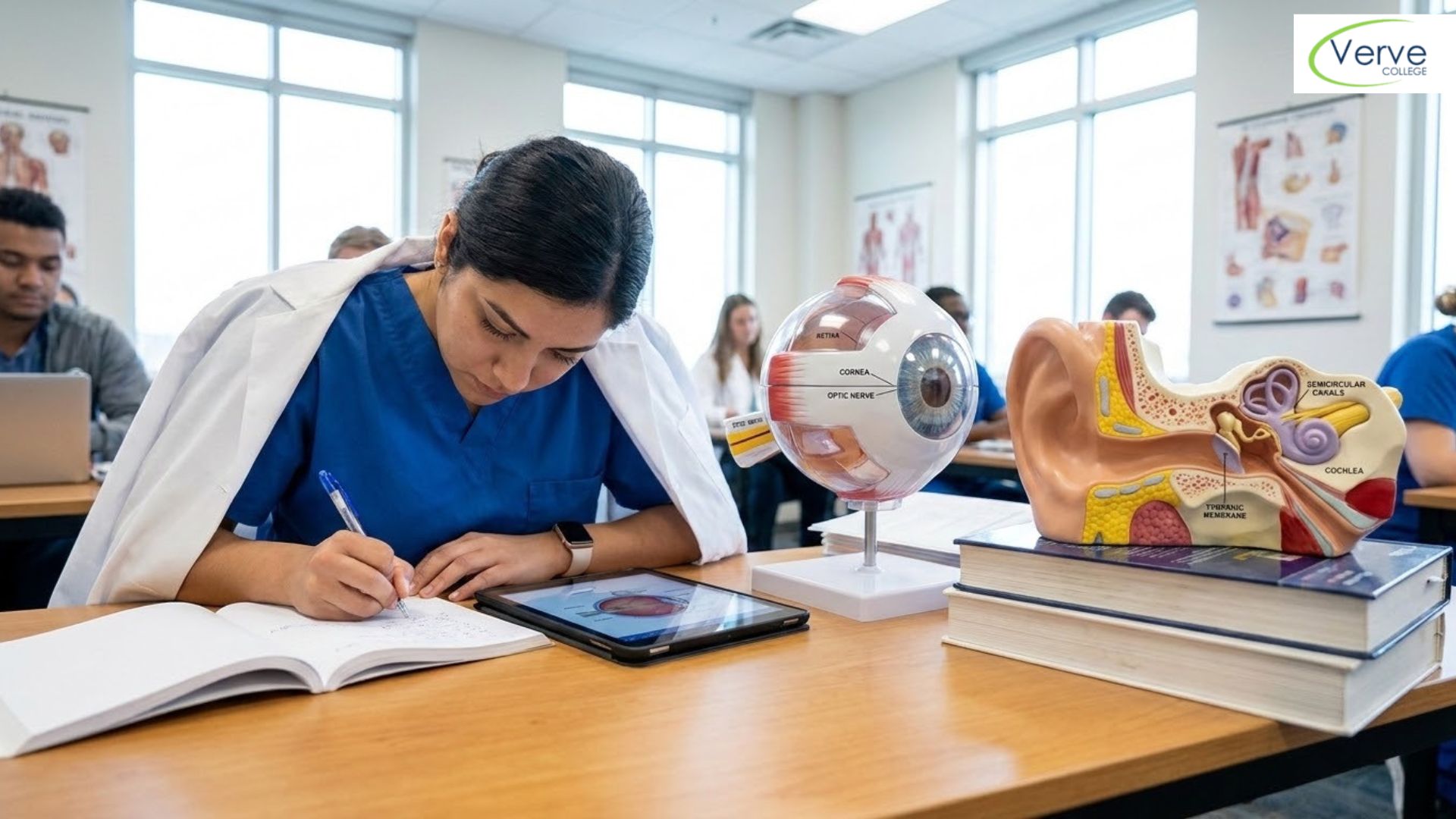
Eye vs Ear Anatomy: Key Differences Every Nursing Student Must Know
Eye vs Ear Anatomy: Key Differences Every Nursing Student Must Know
Many nursing students struggle to remember how different body systems connect and function. The eye and ear are especially confusing because both are sensory organs but work in very different ways. Understanding Eye vs Ear Anatomy is important for exams and real patient care. This guide breaks it down in a simple, practical way so you can actually remember it and apply it in clinical settings.
Key Takeaways
- The eye helps with vision, while the ear controls both hearing and balance
- Each organ has unique structures that serve very specific functions
- The eye works with light, while the ear processes sound waves
- Understanding these differences helps with patient assessment and charting
- If you’re considering an A&P Class, mastering these basics early will make learning easier
What Is Eye Anatomy?
The eye is the organ responsible for vision. It captures light and sends signals to the brain so we can see clearly and respond to our environment.
Key Parts of the Eye
- Cornea: The clear front layer that lets light enter
- Iris: The colored part that controls how much light comes in
- Pupil: The opening that adjusts based on light levels
- Lens: Focuses light onto the retina
- Retina: Converts light into signals for the brain
- Optic nerve: Sends those signals to the brain
How It Works
Light enters through the cornea and passes through the pupil. The lens adjusts to focus that light onto the retina. The retina converts it into electrical signals, which travel through the optic nerve to the brain. The brain then processes these signals into images.
Why It Matters in Nursing
You’ll often assess vision during patient care. For example, checking pupil response can help identify neurological issues. Eye assessments are also important for patients with diabetes or head injuries.
What Is Ear Anatomy?
The ear is responsible for both hearing and balance. It works by detecting sound waves and helping the body stay stable when moving or standing.
Key Parts of the Ear
- Outer ear: Collects sound waves
- Middle ear: Amplifies sound using tiny bones called ossicles
- Inner ear: Converts sound into signals and controls balance
Important Structures
- Cochlea: A spiral-shaped structure that turns sound waves into nerve signals
- Semicircular canals: Detect movement and help maintain balance
- Auditory nerve: Sends sound signals to the brain
How It Works
Sound waves enter the ear canal and hit the eardrum, causing it to vibrate. These vibrations pass through the middle ear bones and reach the cochlea. The cochlea converts them into signals that travel to the brain.
At the same time, the semicircular canals detect head movement and help maintain balance. This is why inner ear issues can cause dizziness.
Why It Matters in Nursing
You may encounter patients with hearing loss, ear infections, or balance problems. Knowing how the ear works helps you recognize symptoms quickly and report them accurately.
Eye vs Ear Anatomy: Key Differences
1. Function
The eye is focused on vision.
The ear handles both hearing and balance.
This makes ear anatomy slightly more complex in terms of function.
2. Type of Stimulus
The eye responds to light.
The ear responds to sound waves and movement.
3. Structure Complexity
The eye has a more straightforward structure.
The ear has three main sections with multiple roles.
4. Brain Connection
Both organs send signals to the brain, but through different pathways:
- Eye uses the optic nerve
- Ear uses the auditory nerve
5. Clinical Symptoms
Eye-related issues include blurred vision, dryness, or sensitivity to light.
Ear-related issues include hearing loss, ringing (tinnitus), or dizziness.
If you’re taking anatomy and physiology classes near me, these comparisons will come up often in both written exams and practical labs.
Why This Comparison Matters for Nursing Students
Understanding these differences is not just about passing exams. It directly impacts patient care.
For example:
- A patient reporting dizziness may have a balance issue linked to the inner ear
- A patient with double vision may need neurological or eye-related evaluation
As a nurse, you are often the first to notice symptoms. Knowing the difference helps you take the right action faster and communicate clearly with the healthcare team.
Common Mistakes Students Make
One common mistake is thinking the ear only helps with hearing. In reality, balance is a major function controlled by the inner ear.
Another mistake is confusing how signals are processed. The eye uses light, while the ear depends on vibrations and motion.
Students also tend to memorize parts without understanding their function. This makes it harder to apply knowledge in real situations.
If you’re searching for anatomy classes near me, focusing on understanding instead of memorizing can make a big difference in long-term success.
How to Study Eye vs Ear Anatomy Effectively
1. Use Side-by-Side Comparison
Studying both systems together helps you see the differences clearly instead of mixing them up.
2. Practice Labeling Diagrams
Repeatedly labeling eye and ear structures helps build memory faster.
3. Use Real-Life Examples
Think about symptoms like blurry vision or dizziness. Connecting anatomy to real situations improves retention.
4. Teach Someone Else
Explaining these concepts to a classmate or even out loud to yourself helps reinforce learning.
Practical Next Steps for Students
If you’re planning a career in nursing, building a strong foundation in anatomy is essential.
- Start with basic concepts before moving to advanced topics
- Focus on understanding how each structure works
- Use structured learning to stay consistent
If you’re feeling unsure where to begin, enrolling in a beginner-friendly program can give you the structure and support you need to stay on track.
Clarification: Why Students Confuse These Systems
Many students confuse the eye and ear because both are sensory organs connected to the brain. But the way they collect and process information is completely different.
The eye deals with light and visual signals, while the ear handles sound and movement. Keeping this one idea clear can help you avoid confusion in exams and clinical scenarios.
Conclusion
The difference between the eye and ear goes beyond just vision and hearing. Each organ has a unique structure, function, and role in patient care. Understanding Eye vs Ear Anatomy helps you think clearly during exams and real clinical situations. The stronger your basics, the easier it becomes to build advanced nursing skills and confidence.
Get Your Nursing Career Training Readiness Score Now!
FAQs
- What is the main difference between eye and ear anatomy?
The eye is responsible for vision and processes light, while the ear handles hearing and balance using sound waves and motion. - Why do nursing students struggle with ear anatomy?
Because it includes multiple parts and functions, especially balance. This makes it harder to remember compared to the eye. - What is the best way to learn anatomy for nursing?
Focus on understanding how each part works, use diagrams, and follow a structured learning approach through a guided program.
Most Popular Blogs Posts

Calcium Homeostasis Explained for Nursing Students
July 27, 2026 Read More
Introduction to Microbiology for Nursing Students
July 27, 2026 Read More
Understanding Blood Clotting Step by Step
July 23, 2026 Read More
How Hormones Keep the Body in…
July 22, 2026 Read More